What determines spatial resolution in radiography?
Spatial resolution in radiography describes the ability of an imaging system to distinguish small, closely spaced structures as separate objects. High spatial resolution allows fine anatomical detail to be seen clearly, while poor spatial resolution causes structures to appear blurred.
Understanding the physics
Spatial resolution depends on how accurately the imaging system preserves the spatial location of X-ray photons detected at the image receptor. Several physical processes can blur this information and therefore reduce image sharpness.
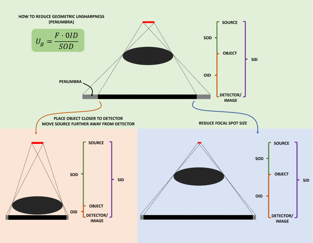
One important factor is the size of the focal spot in the X-ray tube. X-rays are produced from a finite area on the anode rather than from a perfect point source. Because photons originate from slightly different locations within the focal spot, edges in the object produce a small region of partial shadow at the detector known as the penumbra. Larger focal spots produce greater penumbra and therefore reduce spatial resolution.
The degree of geometric blur caused by the focal spot is described by the geometric unsharpness equation:
Ug = (F × OID) / SOD
where Ug is geometric unsharpness, F is focal spot size, OID is object–image distance, and SOD is source–object distance.
This relationship shows that geometric blur increases when the focal spot is larger, when the object is further from the detector, or when the X-ray tube is closer to the patient.
Detector characteristics also influence spatial resolution. Digital radiography images are composed of pixels, and the size of these pixels determines how finely the image can be sampled. Smaller pixels allow finer spatial detail to be recorded.
Detector design can also influence resolution. In indirect detectors, visible light generated in the scintillator may spread slightly before reaching the photodiodes, which can reduce spatial resolution. Structured scintillators such as cesium iodide reduce this effect by guiding light toward the detector elements.
Finally, motion during the exposure can blur the image. If the patient or anatomical structures move while the exposure is being made, the detected signal is spread over a larger region of the detector, reducing image sharpness.
These factors together determine how well the radiographic system can reproduce fine detail.
Where this matters clinically
Spatial resolution is critical when imaging structures where small details must be clearly visualised, such as subtle fractures, fine cortical bone detail, or microcalcifications in mammography.
Radiographic technique can influence spatial resolution. For example, using a smaller focal spot, minimising object–detector distance, and reducing exposure time to limit motion can all improve image sharpness.
However, improving spatial resolution often requires balancing other considerations such as tube loading limits and image noise, so imaging protocols must optimise overall image quality rather than a single parameter.
Related questions
Why does focal spot size affect image sharpness?
What is geometric unsharpness?
Why does increasing SID improve spatial resolution?
What causes motion blur in radiography?