What is geometric unsharpness?
Geometric unsharpness refers to image blur caused by the finite size of the X-ray focal spot and the geometry of the imaging setup. Because X-rays originate from a small area rather than a perfect point source, the edges of structures are projected onto the detector with a region of partial shadow known as the penumbra.
Geometric unsharpness is image blur caused by the finite size of the X-ray focal spot and the geometry of the imaging setup.
This penumbra produces a loss of edge sharpness in the radiographic image.
Understanding the physics
In an ideal imaging system, X-rays would originate from a point source, producing sharp projections of structures onto the detector. In reality, the focal spot on the anode has a finite size. X-rays therefore originate from slightly different positions within the focal spot.
When an object lies in the beam, X-rays from different points of the focal spot project slightly different shadows onto the detector. Instead of a perfectly sharp boundary, a transition zone forms at the edge of the projected structure. This zone is known as the penumbra and appears as edge blur in the image.
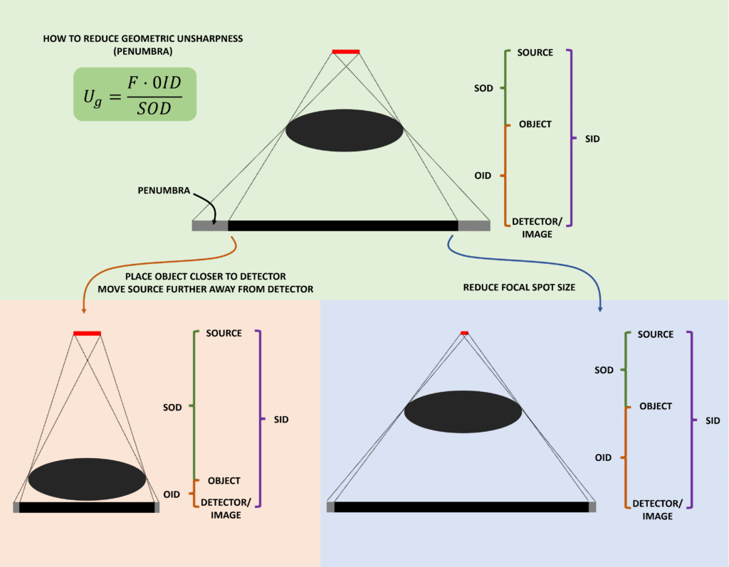
The magnitude of this blur is described by the geometric unsharpness equation:
Ug = (F × OID) / SOD
where Ug is geometric unsharpness, F is focal spot size, OID is object–image distance, and SOD is source–object distance.
This equation shows that geometric unsharpness increases when the focal spot size increases, when the object is further from the detector, or when the X-ray source is closer to the object.
Conversely, geometric unsharpness decreases when the focal spot is small, when the object is placed close to the detector, and when the source-to-object distance is increased.
Where this matters clinically
Geometric unsharpness is an important determinant of spatial resolution in radiography. Imaging techniques aim to minimise geometric blur by optimising the imaging geometry.
In practice, this means using a small focal spot, placing the patient as close as possible to the detector, and increasing the source–image distance (SID) where feasible.
However, these adjustments must be balanced with other considerations. For example, small focal spots limit the allowable tube loading due to heat constraints, and increasing SID may require higher exposure factors to maintain adequate detector signal.
Understanding geometric unsharpness helps explain how radiographic technique influences image sharpness and why imaging geometry is carefully controlled in clinical practice.
Related questions
What determines spatial resolution in radiography?
Why does focal spot size affect image sharpness?
Why does increasing SID improve spatial resolution?
Why does increasing OID degrade image quality?