
Step-by-step mechanism
- Incident photon interaction
- A photon with energy typically between 30–150 keV approaches an atom.
- It interacts with an outer-shell (valence) electron, which is only weakly bound to the atom.
- Energy transfer and electron ejection
- The photon transfers part of its energy to the electron.
- The electron is ejected from the atom as a Compton (recoil) electron.
- The remaining photon energy is reduced and emitted in a new direction.
- Scattered photon emission
- The scattered photon may:
- Undergo further interactions
- Escape the patient (contributing to staff exposure)
- Reach the detector as scatter, reducing image contrast
- The scattered photon may:
Energy conservation: Total energy of incident photon = kinetic energy of ejected electron + energy of scattered photon.
Relationship between scattered photon energy and scatter angle
The energy of the scattered photon depends on its scattering angle (θ):
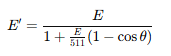
Where:
- E = incident photon energy (keV)
- E′ = energy of the scattered photon (keV)
- 511 keV = rest energy of the electron (mc²)
Key relationships:
- Forward scatter (θ ≈ 0°) → photon retains most of its energy.
- 90° scatter → photon loses moderate energy.
- Backscatter (θ ≈ 180°) → photon energy lowest (~0.2 MeV for 100 keV incident photon).
Energy and Atomic Number Dependence
- Probability roughly proportional to electron density, and therefore to material density (not Z).
- Relatively independent of atomic number (occurs similarly in bone and soft tissue).
- Decreases slowly with increasing photon energy.
- Dominates at higher energies (above ~60–80 keV in tissue) in proportion to the photoelectric effect.
Much like the previous lesson on the photoelectric effect. Let’s place this comparison table here for reference.
Photoelectric effect vs Compton scatter
| Feature | Photoelectric | Compton |
|---|---|---|
| Photon energy | Low | Moderate–high |
| Electron shell | Inner (bound) | Outer (loosely bound) |
| Photon outcome | Completely absorbed | Scattered, lower energy |
| Image effect | Increases contrast | Reduces contrast, increases noise |
| Dose contribution | High (complete absorption) | Moderate |
| Dependence | ∝ Z³/E³ | ∝ 1/E and electron density, ~independent of Z |
| Dominance | Low-energy photons, high-Z materials | High-energy photons, low-Z materials |
Why is this important?
1. Image Degradation
- Scattered photons reaching the detector add unwanted background exposure.
- This reduces image contrast by blurring the distinction between structures.
- Particularly problematic in:
- Large field sizes (abdomen, pelvis, chest).
- Thick body parts (↑ volume of tissue to scatter).
2. Radiation Protection
- Scattered photons are the primary source of occupational exposure for staff and adjacent tissues in the patient.
- Intensity of scatter decreases rapidly with distance: Exposure ∝ 1 / r2
- Standing behind lead barriers or increasing distance from the patient greatly reduces dose.
3. High-kVp Imaging
- At higher tube voltages (>100 kVp), Compton scatter dominates.
- This is desirable for penetration (e.g. chest radiography) but results in lower contrast.
How then do we reduce scatter?
Methods for reducing scatter
| Method | Principle | Effect |
|---|---|---|
| Collimation | Reduces irradiated volume → fewer interactions | ↓ Scatter production |
| Grids | Lead strips absorb angled scatter | ↑ Image contrast |
| Air gap | Increasing distance between patient and detector | Allows scatter to miss detector |
| Compression | Reduces patient thickness (e.g. mammography) | ↓ Scatter generation |
| Lower kVp (when appropriate) | Reduces Compton relative to photoelectric | ↑ Contrast (but ↑ dose because we have to compensate by increasing mA to maintain exposure and more photoelectric effect occurs at lower kVp) |
Key takeaways and exam tips:
- Compton scatter: partial energy transfer between photon and outer-shell electron.
- Scattered photon: lower energy, deflected direction.
- Dominates above ~50 keV in soft tissue.
- Independent of atomic number, depends mainly on electron density and slightly on photon energy.
- Degrades image contrast and contributes to scatter radiation exposure.
- Reduced by: tight collimation, grids, compression, and distance.
- Common exam question: “Describe the Compton effect and explain its significance in diagnostic imaging.”
Up Next
Next, we’ll briefly cover Coherent (Rayleigh) Scatter, a minor but conceptually relevant interaction that contributes minimally to dose or image formation but may appear in exam questions.